By Mathieu Boudreau
The January 2020 MRM Highlights interview is with Zaki Ahmed and Ives Levesque, researchers at McGill University in Montreal, Canada. Their paper is entitled “Pharmacokinetic modeling of dynamic contrast‐enhanced MRI using a reference region and input function tail”. With a new year (and a new editor-in-chief) come new beginnings: as of 2020, MRM Highlights interviews will no longer be selected MRM Editor’s Pick articles. Instead, the MRM Highlights treatment will be reserved for papers that demonstrate reproducible research practices. Accordingly, the above-mentioned paper was chosen because in addition to sharing their code, the authors shared scripts that reproduce every single figure from their article (even the supplementary figures). This interview focuses on the authors and their work, and another blog post, due to be released next week, will provide insights into their reproducible research practices.
MRMH: To start with, could you tell us a little bit about yourselves?
Zaki: I did my undergrad at the University of Windsor where I studied medical physics. Then, for my masters, I came here to McGill where I joined Ives’ lab. I decided to stay with him to do my PhD, and that is what I’m currently working on.
Ives: I first came to McGill as a graduate student. This is also where I studied with Bruce Pike for my masters and did my PhD work in neuroimaging, which focused on white matter imaging. I then did a postdoc at Stanford, where I learned about image reconstruction and 7 Tesla imaging while being advised by John Pauly and Brian Rutt. I came back to Montreal to take up a faculty position in the Medical Physics Unit, where I started the MRI Methods Research group.
MRMH: Could you give us a brief overview of this paper and the work you’ve done here?
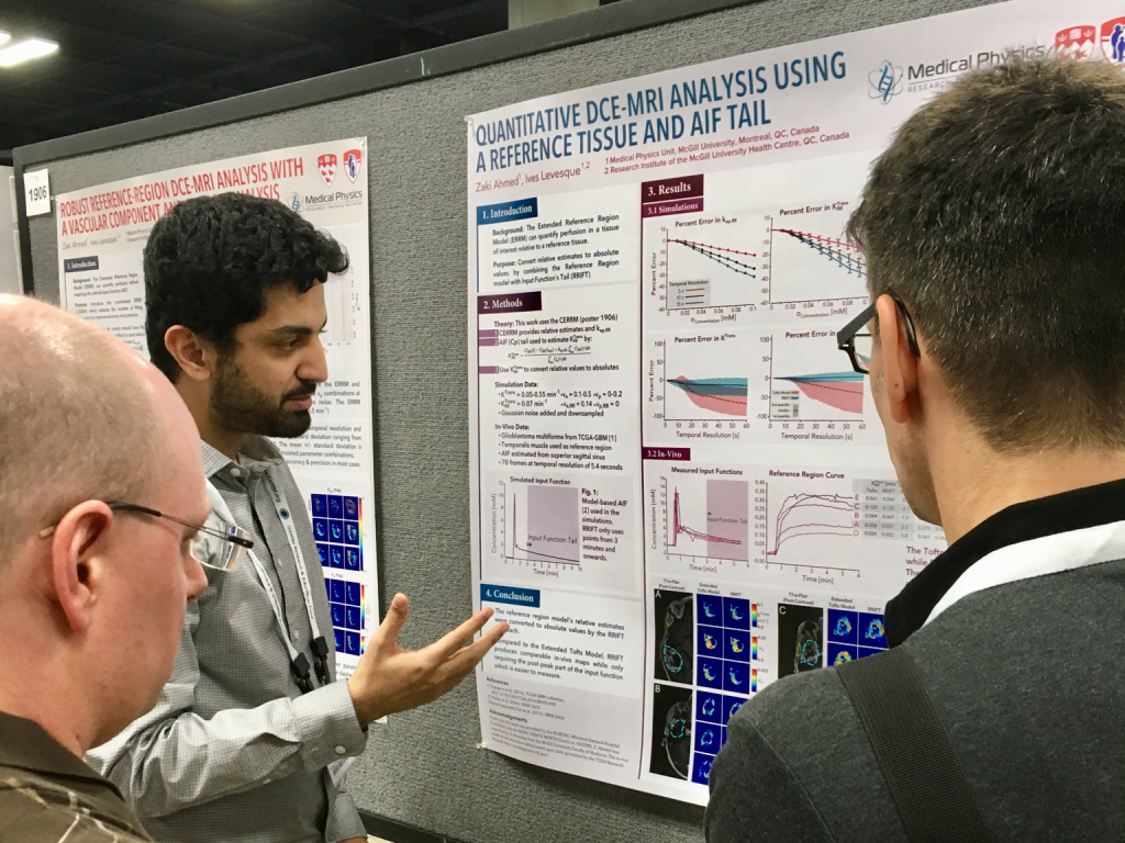
Zaki: So, DCE-MRI involves injection of a tracer followed by a rapid series of scans in which we are able to see the arrival and the uptake of the tracer in tissue. Cancer likes blood and so, thanks to the tracer, we get to see affected regions. But to get quantitative information, we need to know a blood vessel’s contrast-time curve (the arterial input function), which has a very rapid peak followed by a more steady-state tail. To measure the rapid peak, you need a rapid scan, even if this is at the expense of image quality. But it’s a bit of a Catch-22, really, because blood vessels are small, so if you sacrifice image quality it’s hard even to see them. Our work initially focused on the reference region model, in which healthy tissue (e.g. muscle) is used as a reference, but that technique only gives you semi-quantitative results: ratios of quantitative DCE values in the tumor relative to the muscle. So, in this paper, what we proposed was to use the latter part of the input function, the tail (so called because it’s flat), as this would allow us to get away with using a slow scan. One of the benefits of this technique is that even with a slow scan, you still get good fully quantitative maps.
Ives: The first time I spoke about this work to a group outside our university, I was met with little bit of disbelief. For those who understand the quantitative Tofts model and more advanced DCE models, one intuition is that the Ktrans variable (the transfer constant, which is the flow of blood plasma per unit volume of tissue) is strongly determined by the initial part of the enhancement curve. And so the widespread thinking is that you need high temporal resolutions in order to collect those dynamics and measure Ktrans. In reference region modeling, you get Ktrans relative to the reference region. This work is sort of a backdoor calibration method that enables you to get back to the absolute Ktrans by considering the integral of the pseudo steady-state contrast agent concentration. It’s a nice trick, which Zaki thought of, that circumvents the need for temporal resolution while still giving you absolute parameters.
MRMH: Why is this work important?
Zaki: At present, quantitative DCE-MRI is prevalently a research technique, because it requires a fast scan but also high resolution with low noise, and this makes it really difficult to perform clinically, especially in certain cases, like breast tumors. There are some groups that are working on acceleration techniques to make these scans faster, but most clinics don’t yet have access to these. Our technique lets you get these quantitative maps from low temporal resolution data using conventional imaging protocols; this should make them more widely accessible.
Ives: My view is that although the development of fast 4D high-resolution acquisitions will continue to progress in the coming years, at some point the frames are going to be so close in time that SNR may become more of a limitation. This analysis method lets you acquire the necessary images while retaining good SNR.
MRMH: Let’s just touch on the topic of sharing your code. Few people share code along with their papers. This might be, for example, because they’re afraid of getting scooped on future work, or because they feel their code isn’t clean enough to share. Did you experience some of those anxieties before deciding to share your code with your paper?
Zaki: Feeling that your code is not clean enough is pretty par for the course in academia! As regards the code I shared now, I felt that way about several files that I wrote. You have to weigh up the costs and benefits of refactoring a lot of code that might not be essential. As for the fear of being scooped, that was never on my mind. Code sharing is frequent within the DCE-MRI community. In particular, last year at ISMRM there was the Open Source Initiative for Perfusion Imaging (OSIPI), where we got together and discussed good practices for sharing code. I think I feel most anxious when things actually work; that makes me a bit suspicious, because usually they don’t! [chuckles] So my biggest worry is that I may have made a mistake someplace in the code. But even if there is a mistake, it’s better for the work to be shared; that way, it might be found by someone else. Surely, that is better than to publish something that’s wrong but can’t be verified.
MRMH: That’s the way science should work, I would say.
Ives: Yes, I agree with you, it’s the way science should work. You need to have a good dose of humility. What’s more, if you open up your notebook to someone and they say it’s solid, then you have more confidence moving forward.