By Mathieu Boudreau
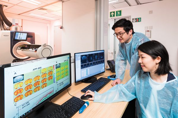
This MRM Highlights Pick interview is with Jianpan Huang and Kannie Chan, researchers at City University of Hong Kong. Their paper is entitled “Deep neural network based CEST and AREX processing: application in imaging a model of Alzheimer’s disease at 3 T”. We chose to highlight this paper because the authors demonstrated exemplary reproducible research practices; in particular, they shared code and sample data in addition to a demo and scripts that reproduce some of their figures.
To discuss this Q&A, please visit our Discourse forum.
MRMH: Tell us about yourselves and your background.
Jianpan: I received my bachelor’s and master’s degrees from Xiamen University, which is where I started getting involved with MRI. I then did my PhD degree in biomedical engineering at the City University of Hong Kong, where I’m currently working as a postdoc. My research focus is CEST MRI and deep learning, and their applications in detecting neurological disease.
Kannie: Actually, I’m a chemist by training, but I’ve always been fascinated by imaging molecules in the body. I went to Johns Hopkins Medicine for my postdoctoral training, which is where I first started working in MRI applications, and then became an assistant professor there. In 2016, I joined the City University of Hong Kong, where I’m now an associate professor. My research focuses on the development of CEST-detectable biomaterial and imaging neuropathology using CEST MRI, mainly for applications in brain cancer and neurodegenerative disease, such as Alzheimer’s disease.
MRMH: Before we dive into the paper, could you briefly explain what CEST and AREX are, for those not familiar with the terms?
Jianpan: So, CEST stands for chemical exchange saturation transfer, and CEST MRI is a technique that probes molecules with exchangeable protons, such as proteins, glucose, and glutamate. AREX is short for apparent exchange-dependent relaxation, which is a relaxation-compensated CEST contrast. It is calculated based on the inverse metric of steady-state Z-spectra and T1 metric, thus correcting the effects of spillover dilution and T1 scaling.
MRMH: Could you give us a brief overview of your paper?
Jianpan: It’s well known that CEST MRI is a promising molecular imaging approach that is sensitive to detect low-concentration molecules with exchangeable protons, however postprocessing CEST data is not trivial in practice and requires expert knowledge to do. It is also quite time intensive. In this study, we used deep learning to process CEST imaging data (deepCEST), an approach that would simplify and accelerate the data processing for users, and applied the technique in a mouse model of Alzheimer’s disease (AD). Moreover, we proposed extension of this deep learning model to predict the AREX contrast (deepAREX). Our results showed that both deepCEST and deepAREX could rapidly generate accurate results at 3T in the mouse model, at higher processing speeds compared with the conventional fitting methods, and the network generalization was validated on unseen data. Interestingly, the deep learning based methods could detect the CEST signal changes in AD mouse brain.
Kannie: By using deep learning, we hope to be able to facilitate the postprocessing, so that it can be shared with others, and that, in turn, will hopefully facilitate clinical translation.
MRMH: Were you surprised with the results?
Jianpan: Yes. The results were quite surprising. A previous study showed that deepCEST could be used to generate CEST contrast, and here we found that deepAREX could also produce high-accuracy AREX contrast that requires additional processing procedures (B0 correction, inverse Z-spectrum analysis, and T1 compensation). Moreover, we were also surprised that the molecular change of AD mouse brain could be detected by CEST MRI (both deepCEST and deepAREX) at clinical-field-strength 3T. This indicates this approach has potential for clinical application.
MRMH: How does this work fit into your broader research goals?
Kannie: We aim to facilitate the clinical translation of CEST using deep learning, as we believe it could be applied in assessing neuropathology, to identify subtle molecular alterations in the brain, for example. We work a lot with animal models because we can perform molecular confirmation or validation to demonstrate the sensitivity, to certain neuropathologies, of the techniques we develop. In another study, we demonstrated that CEST can sensitively detect glucose alteration in Alzheimer’s disease. We really want to make CEST a robust approach that can probe molecular changes in the brain, especially in the earlier stages of the disease.
Jianpan: In this work, we only demonstrated the ability of deep learning to accelerate the postprocessing of CEST MRI data. I’m also interested in applying deep learning to accelerate the acquisition of CEST MRI data. For example, we know that deep learning can be used to reconstruct high-resolution images from low-resolution images, so applying this to CEST MRI could accelerate the data acquisition by just acquiring low-resolution images. Also, there is data redundancy in CEST datasets (in CEST, multiple images from the same location are acquired using different saturation frequency offsets), and deep learning may make it possible to leverage this, through undersampling of the k-space within each CEST image.
MRMH: Since this is a preclinical study, how do you envision the clinical translation of this work?
Jianpan: Reducing the scan time is the first challenge that will need to be overcome, as we just discussed. Lowering the number of saturation offsets is also an option, but at the risk of losing some molecular information. The second challenge is the SAR (specific absorption rate) issues. The approach we used for our animal study was continuous-wave saturation based CEST; however, this is not viable for human studies as it creates high SARs, so a pulsed CEST approach will be used instead. As for the deepCEST and deepAREX, they can be easily translated for clinical usage.
Kannie: Yeah, I definitely agree. The fact that the mouse brain is so different from the human brain means that clinical translation has its challenges, as Jianpan mentioned. Another hurdle is the resolution, because we need to be able to identify very small lesions with our quantitative techniques. We believe emerging deep learning approaches could facilitate clinical translation.
MRMH: You shared some code and data with your paper. What motivated your decision to do so?
Jianpan: There are two main reasons: First of all, we chose to do it for reproducibility purposes. CEST analysis using deep learning is an emerging CEST postprocessing method that is not widely used in this setting. Our hope, in sharing our code/data, is that researchers who are working in the CEST field and/or are interested in deep learning-based CEST analysis might be able to use our code directly for processing their own CEST data. The networks in this study were trained on CEST data acquired from a cohort of mice. Although the networks cannot be used directly to perform prediction tasks in other CEST studies, the shared code could easily be applied for training different CEST datasets, such as human CEST data or CEST data acquired using other CEST parameters.This could make it possible to generate the specific networks for prediction. The second reason is to promote the deep learning based CEST/AREX postprocessing method. We believe that this method would have more impact if it were easier for researchers to use the shared code and apply it to their studies. This could provide an avenue for researchers to share their ideas and thus help to improve this approach.
MRMH: Is there anything else that you’d like to mention?
Kannie: Yes, in addition to what we’ve discussed, I’d like to highlight that the pulse sequence design is also very important for CEST. For clinical applications, alternative sequence designs such as radial readout could also help if applied to CEST MRI.
MRMH: Finally, what do you enjoy doing when you’re not in the lab?
Kannie: I like running outside when I can! Currently we’re under some restrictions again, unfortunately, so I’ve also been enjoying practicing yoga, playing the piano and painting at home.
Jianpan: I enjoy playing basketball quite a lot. Hong Kong is also such a beautiful place, and I like to go hiking or biking if the weather is nice.