By Mathieu Boudreau and Nadia Blostein
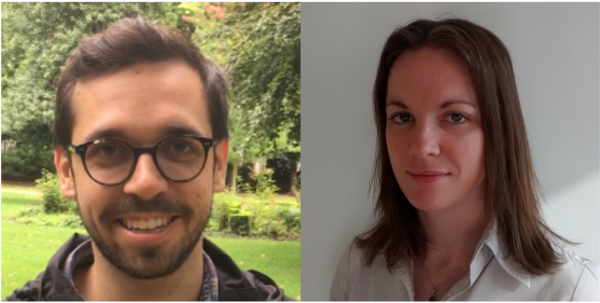
This MRM Highlights Pick interview is with Yaël Balbastre and Martina F. Callaghan, researchers at the Wellcome Centre for Human Neuroimaging at UCL in London. Their paper is entitled “Correcting inter-scan motion artifacts in quantitative R1 mapping at 7T”. Their article was chosen as this month’s Highlights pick because it demonstrated exemplary reproducible research practices; in particular, they shared their code and integrated it in an open-source toolbox (hMRI), as well as shared scripts that reproduces all their simulation figures.
MRMH: Could you tell us about yourselves and how you got into MR?
Yaël: I’m not an MR physicist by training, I studied computer science in France. While I was doing my PhD there, I worked on MR image segmentation in a preclinical imaging context; after that, I moved to London for a postdoc where I worked with John Ashburner and Martina. Martina was the one who somewhat dragged me into the MR physics side [chuckles], and I found it really cool to see how everything works in the image acquisition process. Now I’m currently in Boston working at the Martino Center.
Martina: As for me, I first became aware of MRI as a technique when I was studying physics in Ireland, and thought it was just super cool. I knew I wanted to study it for my PhD, and that took me to London where I worked in Jo Hanjal’s lab in Hammersmith Hospital at Imperial College. After my postdoc there I left MRI for a while, but came back into it about 10 years ago, when I joined the Department of Imaging Neuroscience that hosts the Wellcome Center for Human Neuroimaging. I really enjoy the work environment here, as I get to meet people coming from different backgrounds in computer science and cognitive and computational neuroscience; it’s very diverse.
MRMH: Before jumping into the paper, could you give us a brief explanation of the receive B1 field (B1-) and the transmit B1 field (B1+)?
Yaël: Sure, so the receive field is non-uniform because the sensitivity of the coil elements, used to measure the MR signal, changes according to how far away the source tissue is; the farther you are from the coil elements, the lower the signal will be. When the image is reconstructed, this will show up as darker areas and brighter areas, which represent purely multiplicative signal changes. It is actually the transmit field that rotates the bulk magnetization out of alignment with the main magnetic field, which will then precess and generate a signal in our receiver coils. The longer the transmit field is turned on, the larger the flip will be, and this will, typically, result in a larger signal. But this transmit field is also going to vary proportionally to the distance from the tissue, so you will get different flip angles in different locations in the volume. However, the transmit field has a multiplicative effect on the flip angle, and this will change the signal differently depending on the pulse sequences.
Martina: I’d just like to add that a fundamental assumption in many quantitative MRI techniques is that the receive field variations just cancel each other out. But a few years ago, we realised that this assumption is violated if the patient moves between measurements; this type of motion resulted in low reproducibility in our R1 maps.
MRMH: Could you expand on that? In quantitative MRI, even if the patient moves between measurements, why doesn’t image registration result in the receive fields “cancelling each other out” in any case?
Martina: I think the fundamental point is that, although the person has moved, the coils themselves are static. In other words, since the tissues are at different distances from the coils in the two positions, the receive field will be modulated differently, and image registration can’t correct this difference in the images.
MRMH: Could give us a brief overview of the paper?
Yaël: The paper is concerned with inter-scan motion in quantitative MRI, where you have to acquire multiple scans with different parameters. For the R1 mapping technique we were interested in, just two acquisitions are needed. As Martina said, this type of motion will lead to different receive field modulations between the two positions. In a previous paper, Martina suggested how these modulations might be corrected at 3T by using the body coil, but at 7T this isn’t possible because there is no body coil, so our aim with this application was to build on her previous work. We figured out that if the relative field between the two positions is known, you don’t need to actually measure both receiver fields to correct for them sufficiently for qMRI. So, the main point of the paper is that you can correct for these modulation changes in qMRI by acquiring rapid images before your main scans only from the array coil, in order to estimate the relative sensitivity between two positions. We also found that while the transmit field is “flat” enough to do this simple relative receive field estimation at 3T, at 7T this is not true, so we improved on our understanding of the correction for this case.
Martina: Yes, the transmit field changes at ultra-high fields impact the calibration data themselves, which then become transmit field sensitive. And that was the part that we explored with our simulations. Although our correction works well, we still have more to do in order to address this transmit field issue.
MRMH: What prompted your decision to share code and data with your paper?
Yaël: We always try to share code when we can, and since the hMRI toolbox we used for R1 processing is open-source, it made sense to share it there too. As for the simulations, that was all Martina’s doing.
Martina: It’s also important to note that the institute where I am working, and where Yaël was at the time of the study, is the home of SPM. And so, we’ve been surrounded by the whole open-source ethos that was adopted here since the 90s, that is, since before open science was really a thing. With the hMRI toolbox, too, the idea was to make it all available. More recently, we’ve tried to do the same for simulations used in our papers and make them available as well. It’s beneficial because if somebody wants to really learn how you did something, this really makes it easier for them to understand your work. From my perspective, I started doing research when none of these tools like GitHub existed, so I’ve really benefited from the trainees joining the lab, and those that are part of the hMRI development consortium, showing us what’s new and can potentially boost the impact and reproducibility of our research.
MRMH: To finish off, what do you enjoy doing when you’re not in the lab?
Martina: Well, I loved it when Yaël was here, because Yaël, Nadège (she’s also a co-author on the paper) and I would play board games and card games a lot. Those two were really into puzzles, and it was just lovely to see how they would never be put off trying to figure out whatever the problem was, and I could then see them applying that same approach to their work. I also really love going to gigs as well; the last one I went to was Metronomy, who were playing in London during ISMRM.
Yaël: At the moment, I’m just trying to make the most of being in the US. So, I’ve been visiting a lot of parks and such. Last weekend I went for a two-hour drive to go leaf peeping, because the Fall is beautiful here; everything is red, yellow, or orange.